|
| OBJECTIVES | ||||
| At the end of this course, the student will be able to: |
||||
| Describe the basic functions of the circulatory and nervous systems. |
||||
| Name the major divisions of the brain and their functions. |
||||
| List at least five risk factors for stroke. |
||||
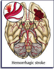
| Describe the difference between hemorrhagic and ischemic strokes. |
||||
| List five signs and/or symptoms of stroke. |
||||
| Describe the treatment for a patient who has suffered a stroke. |
||||
| CASE SCENARIO | ||
 Responder Tip |
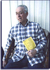
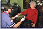
Your "shift" has been called to a private residence for woman who says that her husband is unable to speak. The woman, who identifies herself as Mrs. Jones, greets you at the door. She tells you that her husband was on the porch reading a magazine. When she called him to come inside for lunch, he didn't answer. Concerned, she went out and found him sitting in a chair, awake but unable to speak (aphasia). Your initial assessment of Mr. Jones reveals an elderly man, alert to verbal stimuli. He attempts to speak, but his words are difficult to understand. The right side of his mouth has an obvious droop and he is drooling. He is right handed, but you find his motor strength to be weak on the right. What is your primary concern for Mr. Jones? What action will you take first in his immediate care?
|
|
| INTRODUCTION | ||
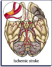
| Stroke is the third leading cause of death in this country and accounts for more than 150,000 deaths annually. Just as a heart attack can be caused by compromise in the coronary arteries, stroke can be caused by a compromise in the arteries supplying the brain. The common risk factors for stroke have been clearly identified, as well as the common signs and symptoms seen in most stroke patients. Definitive care for the stroke victim depends on the type of stroke, but prehospital care is largely the same for all stroke patients.
|
| ANATOMY AND PHYSIOLOGY OF THE CIRCULATORY SYSTEM | ||
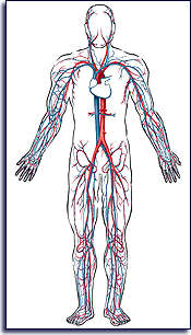 Arteries (red) and veins (blue). |
The circulatory system is a complex transportation network designed to deliver oxygenated blood to every cell in the body. The vessels that channel the blood from the heart to the body's tissues are called arteries. Arteries branch into smaller vessels called arterioles. Vessels that return blood from the tissues to the heart are called veins. Small veins are called venules. The tiny vessels that carry blood through the tissues are called capillaries.
Blood Vessels and Circulation Functionally, capillaries are the most important. Capillaries carry out a two-way exchange of fluid, nutrients, electrolytes, hormones, waste products, and other substances between the blood and tissue cells. The network of capillaries in the tissues and organs is called the capillary bed. The flow of blood through the capillary bed is called microcirculation. Blood pumped out of the heart flows into the arteries. Since the arteries transport blood under high pressure, they have strong walls that facilitate rapid blood flow. Major arteries distribute the blood to different body regions before branching into distributing arteries, which lead to specific organs and muscles. Most arteries carry oxygen-rich blood from the heart to the body's organs. It is the pulmonary arteries that are an exception to this rule. They carry unoxygenated blood from the heart to the lungs for oxygenation, and yet are still referred to as arteries due to the fact that they carry blood away from the heart.
|
|
 The left and right coronary arteries provide blood supply to the heart. |
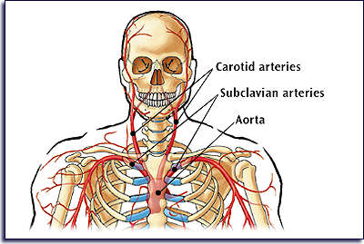
The aorta is the main artery leaving the heart. It has several sections from which the major systemic arteries branch off to supply various body regions. The left and right coronary arteries are the first major blood vessels to branch off from the aorta as it leaves the left ventricle. Cardiac muscle tissue requires a constant supply of oxygenated blood, which is provided by the coronary artery system. Any interruption of the blood supply to the heart will quickly result in injury and death of the myocardium, affecting its ability to pump blood.
The next major branches from the aorta are the carotid and subclavian arteries. The left and right carotid arteries are the major arteries of the neck. They branch to supply blood to the throat, head, brain, and face. Pulsations of the carotid arteries are felt on either side of the neck, just below the angle of the jaw. The subclavian arteries are located directly beneath the clavicle.
Circulation is the rhythmic cycling of blood through the vessels and tissues, driven by the continuous pumping of the heart. The amount of blood pumped by the heart into the vessels each minute is called the cardiac output. This amount varies with the body's requirements. The brain requires a constant blood flow to supply its delicate tissues with oxygen and glucose.
Blood Pressure Often, the stroke patient will have a significantly elevated blood pressure. It is currently believed that the blood pressure rises in an attempt to perfuse the tissues beyond an obstructed cerebral artery.
|
|
| Responder
Tip Systemic hypoperfusion is commonly referred to as shock. |
Perfusion and Hypoperfusion Perfusion refers to the microcirculation of oxygen rich blood through all the organs and tissues. As blood circulates, oxygen and other nutrients are delivered to the cells and waste products are removed. With proper circulation, perfusion is complete, and the nutritional and energy needs of each cell are met. However, if the circulation is inadequate for any reason, the tissues do not receive enough oxygenated blood. This state is called hypoperfusion. Inadequate perfusion leads to hypoxia (oxygen deficiency) and cell death. Cell death is particularly devastating in the brain since nervous system tissue cannot be regenerated.
|
| ANATOMY AND PHYSIOLOLGY OF THE NERVOUS SYSTEM | ||
| The nervous system controls the voluntary and involuntary activity of the body. It senses changes within the body and in the outside environment. The nervous system analyzes all the input, then integrates this information to formulate a response by the body.
Neurons are specialized nerve cells, capable of transmitting electrical impulses. Neurons can relay information over distances as short as one millimeter, or as long as a meter or more. For example, a single neuron carries sensory information from the toes to the base of the brain. Information from sensory receptors throughout the body is carried to the spinal cord and brain by sensory nerves. Outgoing impulses that cause muscles to contract or glands to function travel through motor nerves.
|
||
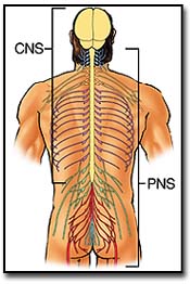 The two divisions of the nervous system are the CNS and the PNS.
|
The Central and Peripheral Nervous System
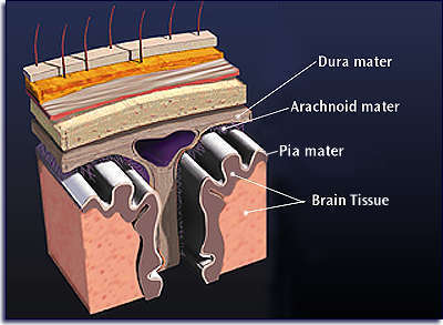
The nervous system has two anatomical divisions, the central nervous system (CNS) and the peripheral nervous system (PNS). The central nervous system includes the brain and spinal cord. The CNS integrates all sensory input and motor output exchanges with the peripheral nervous system. It is the headquarters for memory, emotion, and intellect. The central nervous system is encased in membranes called the meninges. Starting with the deepest layers and moving outward, the layers of the meninges are pia mater, arachnoid mater, and dura mater. A cushion of cerebrospinal fluid bathes and protects the spinal cord and brain. An intracerebral hemorrhage may cause bleeding that can accumulate between the layers of the meninges.
The peripheral nervous system consists of long nerve fibers that carry information between the CNS and the muscles and organs. The PNS carries impulses between the central nervous system and various receptors, muscles, and glands in the extremities and internal organs. Sensory nerves transport information from the body to the brain and spinal cord. Motor nerves carry information from the brain and spinal cord out to the body's structures. A cerebrovascular accident (CVA) occurs in the CNS but often shows its affects through signs and symptoms related to the PNS.
The Somatic and Autonomic Nervous System The nervous system can also be divided according to function. The somatic nervous system (SNS) is involved in conscious perception of the environment and voluntary movements of the body. The autonomic nervous system (ANS) senses the body's internal environment. This system controls involuntary functions such as heart rate, gastrointestinal function, and glandular secretions. The ANS controls the involuntary actions of cardiac muscle, smooth muscle, and glands. Autonomic sensory nerves monitor internal body conditions such as oxygen and carbon dioxide levels in the blood. They also detect the presence of food in the digestive tract. The motor portion of the ANS is divided into sympathetic and parasympathetic systems. These systems have opposite effects upon the smooth muscle, cardiac muscle, and glands that they control.
Stress-related Physiological Changes
|
| STRESS-RELATED PHYSIOLOGICAL CHANGES |
||
|
||
|
||
|
||
|
||
|
||
|
||
|
||
|
| Responder
Tip Stroke patients will frequently have difficulty swallowing making them more prone to airway compromise. Continuous assessment of the airway is essential. |
As you study the signs and symptoms of stroke, notice that many are related to autonomic nervous system functions.
The parasympathetic division of the ANS is responsible for maintaining normal body functions. This system also restores and conserves energy during recovery periods. Parasympathetic activity tends to slow the heartbeat. It enhances smooth muscle contractions for activities such as glandular secretion, the digestion and absorption of food, and the elimination of wastes. It is sometimes called the rest and repair system. Intense fear can increase the activity of this system so much so that one loses control over bodily functions.
The Brain
|
||||
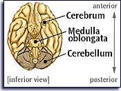 Subdivisions of the brain. |
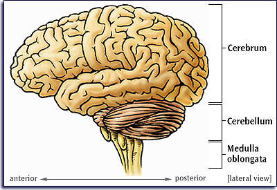 Subdivisions of the brain.
|
||||
|
The cerebrum makes up most of the brain mass. It controls all voluntary movement, stores memory, and is the seat of intelligence, reasoning, and creativity. A deep sagittal groove partially separates the cerebrum into left and right cerebral hemispheres. The hemispheres of the brain have anatomical and functional differences. The right side of the brain controls the left side of the body. Likewise, the left side of the brain controls the right side of the body. For most people, the left side of the brain is important for speech, math, scientific skills, and reasoning. The right side of the brain is involved in spatial and pattern recognition, insight, and creativity. An injury on one side of the brain usually presents with deficits on the opposite, or contralateral, side of the body.
The cerebellum is in the posterior and inferior aspect of the cranium. It helps maintain posture and balance, and governs coordinated, skilled movements such as running or speaking. The medulla oblongata is the section of the lower brain known as the brain stem. It exerts control over autonomic (involuntary) functions. The medulla regulates blood pressure, body temperature, hunger and thirst, sleep and waking cycles, and respiration.
|
| RISK FACTORS FOR STROKE |
||
|
||
|
||
|
||
|
||
|
||
|
||
|
||
|
||
|
||
|
||
|
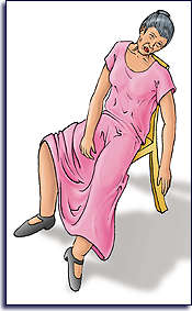 Some symptoms of stroke.
Responder Tip |
Signs and Symptoms of Stroke
Strokes are most commonly seen in elderly patients with a history of hypertension. Other conditions that cause atherosclerosis, such as high cholesterol or diabetes, can also cause strokes. Occasionally, younger people do develop stroke. Signs and symptoms of CVA may include:
|
|
| Responder
Tip TIAs are sometimes referred to as mini strokes.
|
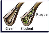
Transient Ischemic Attack (TIA) Another condition similar to stroke commonly seen during EMS responses is the transient ischemic attack (TIA). This may also be called a mini stroke, and may be caused by a temporary embolus or clot that dissolves over time. During a TIA, the patient has symptoms of a stroke but recovers within a short time, usually a few minutes. Occasionally, recovery may take up to 24 hours. Although the patient may recover before transport, a physician should evaluate the patient's condition. Patients with TIA are at high risk for developing a major stroke. Up to 40% of all patients who suffer TIAs will later have significant strokes. Frequently, patients who have suffered TIAs have blockages of the carotid arteries. Surgical removal of fatty deposits in the carotid arteries, called plaques, may be required to correct the condition.
Emergency Care of the Stroke Patient
|
| EMERGENCY CARE OF THE STROKE PATIENT |
||
 If the patient is conscious, conduct a focused history and physical exam. |
|
|
| SCENARIO WRAP UP | ||
| Responder
Tip The patient’s level of consciousness is often your best indication as to their immediate condition. |
As you learn more about this patient from his wife, you discover that he has a history of hypertension and is under considerable stress due to family problems at home. He has been non-compliant with his blood pressure medications and continues to smoke heavily, despite warnings from his physician. You realize that you cannot be sure of the exact cause of his condition, and you act quickly to administer oxygen and complete packaging for transport.
While waiting for EMS, you continue to assess his vital signs, noting his blood pressure to be elevated. Administration of oxygen does not improve his condition. He continues to worsen, developing complete paralysis on his right side. You radio
the patient's condition to the responding EMS unit. The accuracy of your assessment and report enable doctors to start this patient on life-saving medication soon after
his arrival at the Emergency Department.
|
| ACRONYMS FOR THIS COURSE |
||
|
||
|
||
|
||
|
||
|
||
|
||
|
|
Key Terms:
Care of the Stroke Patient (click on a term to hear it pronounced)
|
|||
|
Term
|
Definition
|
||
| Aphasia | inability to speak | ||
| Atherosclerosis | a disease process characterized by a build up of fat, cholesterol, and calcium within the lining of a blood vessel | ||
| Carotid arteries | second major branch from the aorta; the carotid arteries supply blood to the throat, head, brain, and face | ||
| Central Nervous System (CNS) | the brain and spinal cord | ||
| Cerebellum | portion of the brain that coordinates voluntary muscular movements | ||
| Cerebrovascular Accident (CVA) | an acute neurologic deficit that results when an artery in the brain is blocked or ruptured and lasts more than 24 hours. Commonly known as a stroke or more recently brain attack | ||
| Cerebrum | the largest part of the brain separated into two hemispheres (right and left) by a deep sagittal groove | ||
| Contralateral | on the opposite lateral side of the body | ||
| Hemorrhagic stroke | A stroke caused by rupture of a blood vessel and bleeding into the brain or adjacent space | ||
| Hypertension | abnormally high blood pressure | ||
| Hypoperfusion | inadequate perfusion of the cells with oxygen and nutrients resulting in a buildup of metabolic waste products in the tissues | ||
| Medulla oblongata | the widening continuation of the spinal cord, which forms the lowest part of the brain and contains nerve centers that control breathing, circulation, and other important functions | ||
| Neuron | specialized nerve cells capable of transmitting electrical impulses that carry sensory information from the organs to the brain | ||
| Parasympathetic Nervous System | the division of the autonomic nervous system which turns off the "fight or flight" response and turns on vegetative processes such as digestion of food. | ||
| Perfusion | microcirculation of blood within the organs and tissues; involves delivery of oxygen and nutrients to the cells and removal of waste products | ||
| Peripheral Nervous System (PNS) | part of the nervous system not including the brain and the spinal cord; the nerves and ganglia | ||
| Subclavian arteries | arteries beneath the clavicles which deliver blood to the upper portion of the body and to the brain via the right carotid artery, a branch of the right subclavian artery | ||
| Sympathetic Nervous System | division of the autonomic nervous system (ANS) concerned with processes of energy utilization and the "fight or flight" response | ||
| Thrombolytic Therapy | Administering intravenous medications that break down a blood clot to restore perfusion | ||
| Transient Ischemic Attack (TIA) | an acute, focal neurologic deficit that resolves within 24 hours. Commonly called a "mini-stroke" | ||